
Most Beautiful Ramadan Square Competition 2

Under the patronage of
Professor Dr. Ahmed El-Menshawy, President of the University
Professor Dr. Ahmed Abdel-Mawla, Vice President for Education and Student Affairs
Professor Dr. Enas Ahmed Abdel-Hafez, Dean of the Faculty
Professor Dr. Khaled Mohamed Ahmed Hassanin, Vice Dean for Education and Student Affairs
Professor Dr. Madiha Darwish, General Coordinator of Student Activities at the University
And under the supervision of Dr. Ahmed Thabet Abdel-Awad, Director of the Student Welfare Department
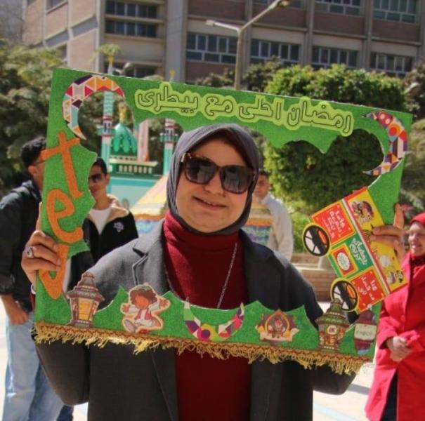
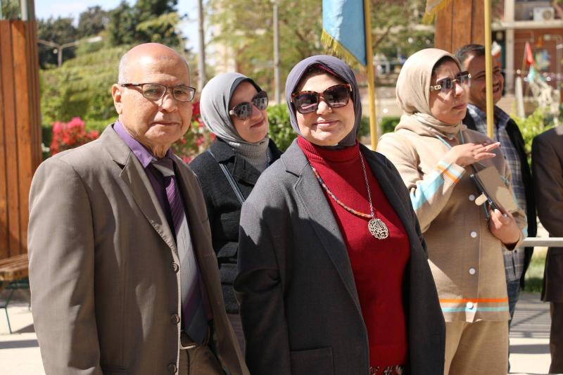
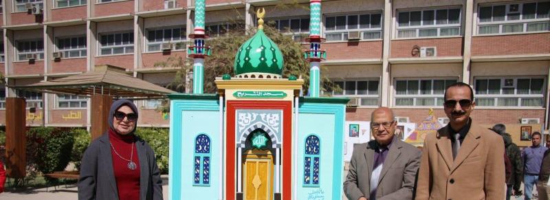
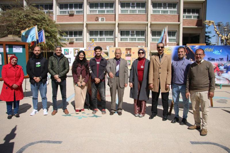
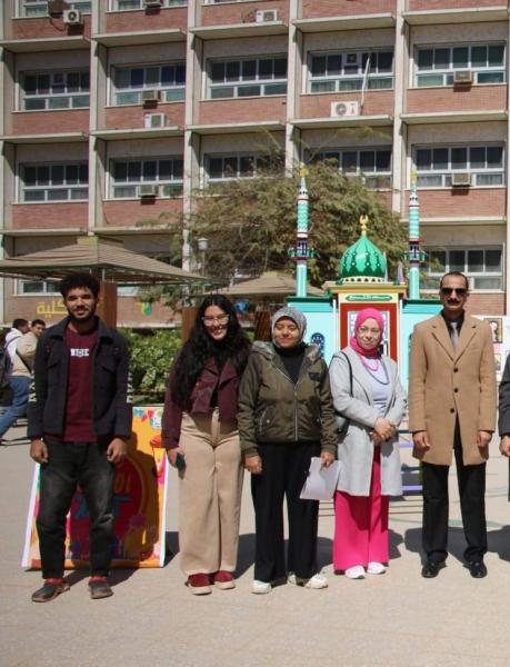
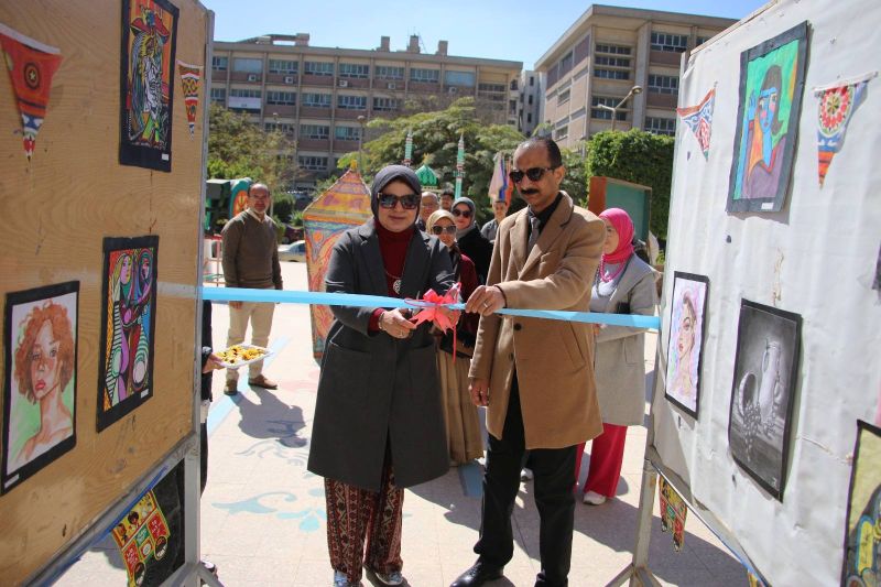
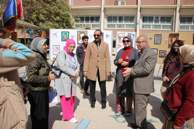
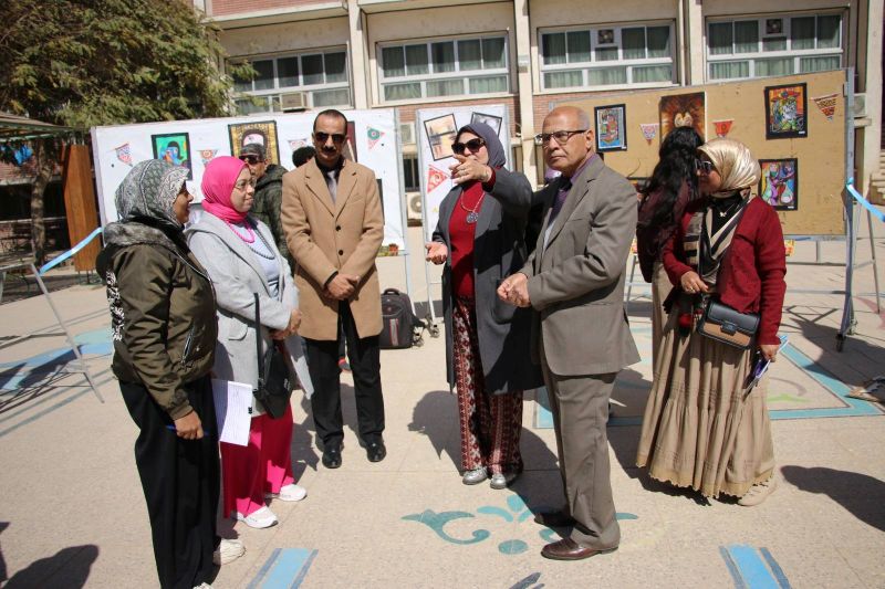
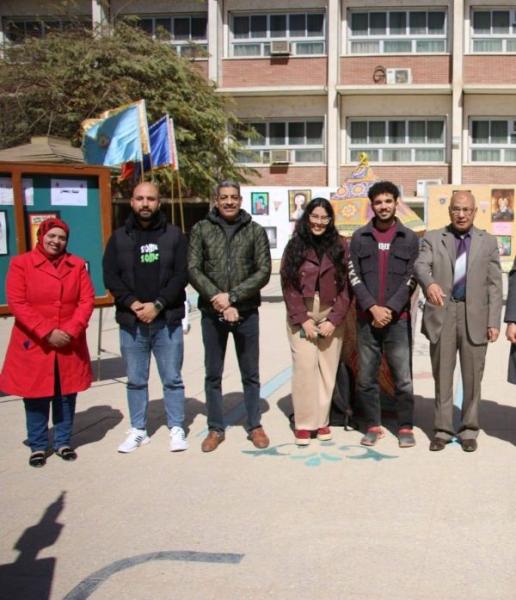
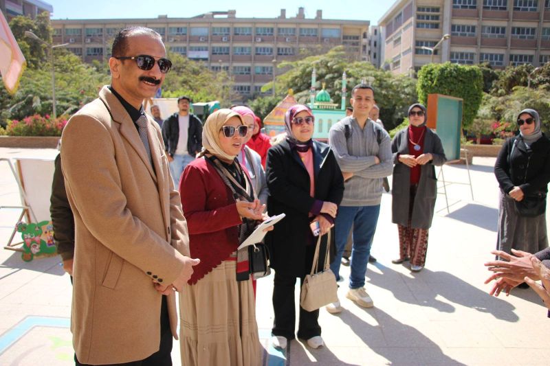
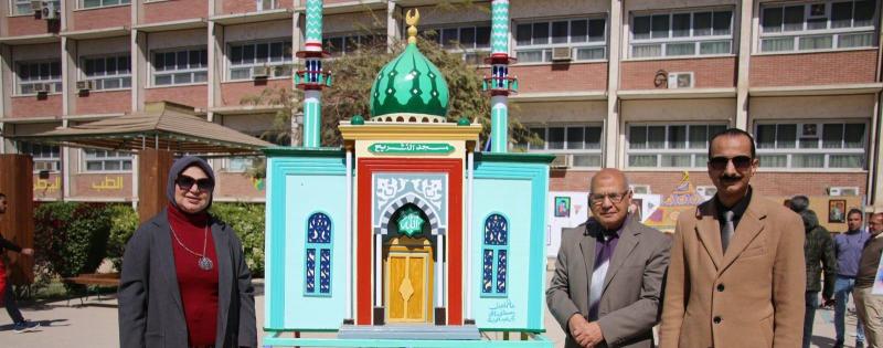
The Technical Committee of the Student Welfare Department at the Faculty of Veterinary Medicine participated in the "Most Beautiful Ramadan Courtyard 2" competition. The evaluation was attended by Professor Dr. [Name of Dean], Professor Dr. [Name of Vice Dean], Professor Dr. [Name of Vice Dean], Professor Dr. Mohamed Ahmed Alam El-Din, Supervisor of the Anatomy Department Museums, and an evaluation committee from the Arts Department of the General Administration for Student Welfare at Assiut University, on Sunday, March 1, 2026.

All praised the high standard of the Ramadan displays in the courtyard of the Faculty of Veterinary Medicine.

1

Art exhibition by student Mohamed Refaat (fourth year) and student Jessica Raafat Fayek (first year)

0

Under the patronage of Professor Dr. Ahmed El-Menshawy, President of the University; Professor Dr. Ahmed Abdel-Mawla, Vice President for Education and Student Affairs; Professor Dr. Enas Ahmed Abdel-Hafez, Dean of the Faculty; Professor Dr. Khaled Mohamed Ahmed Hassanin, Vice Dean for Education and Student Affairs; and Professor Dr. Madiha Darwish, General Coordinator of Student Activities at the University; and under the supervision of Dr. Ahmed Thabet Abdel-Awad, Director of the Student Welfare Department, the Technical Committee of the Student Welfare Department at the Faculty of Veterinary Medicine organized an art exhibition for student Mohamed Refaat (fourth year) and student Jessica Raafat Fayek (first year). The exhibition was inaugurated by Professor Dr. the Dean of the Faculty and Professor Dr. the Vice Dean for Education and Student Affairs, in the presence of a select group of supervisors of fine arts from the Arts Department of the General Administration for Student Welfare at Assiut University. The exhibition opened on Sunday, March 1, 2026, and continued until Tuesday, March 3, 2026, in the Faculty grounds.

All attendees praised the high quality of the artwork. The art in the exhibition

215

Light and Electron Microscopic Characterization of Vascular and Mitochondrial Structures in the Camel Retina

Research Abstract

Camels have unique morphological traits that enable them to adapt well to harsh conditions. This work aims to describe the vascular architecture of the camel retina and investigate its cellular components with a focus on the distribution of mitochondria in Muller cells and photoreceptors, using light and electron microscopy. The camel retina is euangiotic in which blood vessels extend in the inner retina from the nerve fiber layer to the outer plexiform layer. The pericytes are embedded in the basement membrane of the retinal capillaries, and overlapping of pericytes could be observed. Glial cells are localized in the vicinity of blood vessels. Muller cells display mitochondria throughout their length, from their end-feet, which form the inner limiting membrane, to their scleral end, which forms the outer limiting membrane. Interestingly, the bodies of camel Muller cells are densely packed with mitochondria, while their end-feet show few mitochondria. Numerous mitochondria could be observed in the axons and synaptic terminals of rods and cones. Photoreceptor bodies are devoid of mitochondria. The inner segment's ellipsoid region is densely packed with mitochondria, whereas the outer segment lacks them. In conclusion, these findings provide new insights into the vascular and cellular organization of the camel retina, highlighting key adaptations such as a well-developed inner retinal vasculature, specialized features of the inner blood–retinal barrier, and a distinctive pattern of mitochondrial distribution in Muller cells and photoreceptors. This structural specialization may play a crucial role in maintaining retinal function under the challenging environmental conditions camels face.

Research Authors
Manal T Hussein, Wafaa Gaber, Khaled H Aly, Fatma M Abdel‐Maksoud
Research Journal
Microscopy Research and Technique
Research Publisher
John Wiley & Sons, Inc.
Research Website
https://analyticalsciencejournals.onlinelibrary.wiley.com/doi/abs/10.1002/jemt.70073
Research Year
2025

Mizolastine-loaded spanlastics for treatment of atopic dermatitis: immunomodulatory approach for management of inflammation

Research Abstract

Atopic dermatitis (AD) is the most widespread chronic inflammatory skin disease characterised by impaired skin barrier, higher immunoglobulin E (IgE)-mediated sensitisation, and pronounced inflammatory and immune activity. In this study, mizolastine-loaded spanlastics (MZSPs) with edge activators (Brij 35, Tween 80, and Cremophor RH40) were formulated using ethanol injection method. Spanlastics were assessed for their average particle size, surface charge, encapsulation efficacy, morphology, in vitro drug release, and ex vivo skin permeation. MZSPs showed average particle size ranging from 186.2 ± 13.0 nm to 380.7 ± 25.9 nm and high MZ encapsulation efficiency percentage of (95.8 ± 0.68%–98.1 ± 0.86%). The prepared MZSPs were incorporated into 2.5% HPMC hydrogel base to facilitate its topical delivery. The in vitro release experiments showed a sustained release profile of MZ from MZSPs-hydrogel over 24 h with effective flux and permeation through rat skin compared to free drug. Additionally, the therapeutic efficacy of MZSPs-hydrogel in an AD model utilising seven-week-old male Balb/C mice outperformed the free drug in improving cytokine profiles, histopathological and immunohistochemical parameters, along with a quantitative analysis of mast cell immunostaining. These findings support the potential application of MZSPs for the transdermal delivery of MZ, offering enhanced management for AD.

Research Authors
Marwa G Elnaggar, Aml I Mekkawy, Amira F Taha, Fatma M Abdel-Maksoud, Mohamed G Elnaggar, Hesham M Tawfeek, Hassan Tamam
Research Date
Research Journal
Journal of Drug Targeting
Research Member
Research Pages
1-11
Research Publisher
Taylor & Francis
Research Website
https://www.tandfonline.com/doi/abs/10.1080/1061186X.2025.2593462
Research Year
2025

Histopathological effects of lactic acid bacteria enriched white soft cheese in testes of albino rats

Research Abstract

Soft cheeses provide an ideal matrix for bacterial growth, serving as a medium for inoculating starter microorganisms like Lactobacillus. This study aimed to evaluate the biochemical, histopathological, and immunohistochemical effects of white soft cheese inoculated with Lactobacillus helveticus, Lactobacillus rhamnosus, and Streptococcus thermophilus S3855 on male reproductive health in albino rats. Forty male Albino rats, divided into five isolated groups, and fed white soft cheese prepared with different bacterial strains or non-inoculated cheese (control) for 28 days. The prepared cheese was pickled for four weeks at refrigerator temperature and analyzed for chemical and microbiological properties at 7, 14, 21, and 28 days. Total solids decreased over time, while protein-to-dry matter ratios increased across all treatments. Biochemically, all treated groups showed reduced serum total cholesterol, with the most significant triglyceride reduction in group V. Histopathological analysis revealed notable structural damage in testicular tissue, including degeneration of seminiferous tubules, vascular abnormalities, and a marked decrease in seminiferous tubule diameter and spermatogenic cell counts. Johnsen-like scores were significantly lower, particularly in groups III, IV, and V, indicating impaired spermatogenesis compared with the normal control. Immunohistochemically, strong Bax expression was observed in all treated groups, indicating enhanced apoptotic activity. Additionally, wide spread Plexin-B1 expression was also detected in all treated groups. These findings indicate that probiotic-enriched soft cheese may negatively affect male reproductive health, possibly through apoptosis-mediated pathways.

Research Authors
Ruwaida Elhanbaly, Khaled H Salman, Fatma M Abdel-Maksoud, Fatma Abo Zakaib Ali
Research Date
Research Journal
Scientific Reports
Research Publisher
Nature Publishing Group UK
Research Vol
15
Research Website
https://www.nature.com/articles/s41598-025-26073-2
Research Year
2025

Dynamics of the posthatching testicular development in Japanese quail (Coturnix coturnix japonica): histological and ultrastructural study

Research Abstract

The posthatching development of the testis is a well-organized process comprising the maturation of Sertoli cells, the development of Leydig cells, and the differentiation of germ cells. This study aimed to investigate the posthatching testicular development in the Japanese quail, using light and electron microscope. The current study was performed on 25 healthy Japanese quail chicks at 0, 7, 21, 40, and 50 posthatching days. The results revealed that the testis consists of solid seminiferous cords, and their lining epithelium is composed of two types of cells; immature Sertoli cells and gonocyte or spermatogonia at the early stage, which begins from the day of hatching till 21 days posthatching. The interstitium during this period consisted of different developmental stages of the Leydig cells. However, at the late posthatching developmental stage the testis is characterized by the presence of round and elongated spermatids as well as the initiation of spermiogenesis. The interstitial compartment also showed an increase in the number and size of Leydig cells. The findings of the current study provide comprehensive insights into the posthatching development of the Japanese quail testis, contributing to the understanding of avian reproduction.

Research Authors
Sara MM Eldesoky, Marwa M Hussein, Fatma M Abdel-Maksoud
Research Date
Research Journal
Microscopy and Microanalysis
Research Pages
ozaf012
Research Publisher
Oxford University Press
Research Vol
31
Research Website
https://academic.oup.com/mam/article-abstract/31/2/ozaf012/8113782
Research Year
2025

Protective efficacy and immune response to adjuvanted Pseudomonas putida ghost vaccine in Nile tilapia

Research Abstract

Aquaculture faces significant challenges from bacterial pathogens such as Pseudomonas putida, which causes severe infections in Nile tilapia (Oreochromis niloticus), leading to economic losses and public health concerns. To address this, a bacterial ghost vaccine of P. putida (PPGs) was developed using a sponge-like reduced protocol, ensuring structural integrity and complete inactivation while preserving immunogenic surface components. The vaccine was formulated with a water-in-oil-in-water (W/O/W) adjuvant to enhance immunogenicity. Efficacy of the vaccine were evaluated in tilapia through serum IgM quantification, gene expression analysis, and challenge trials. Results demonstrated that both plain and adjuvanted PPGs vaccines elicited robust humoral and cellular immune responses, with significant upregulation of immune-related genes (IL-1β, IL-8, IgM, MHC-II α) and high serum IgM levels. The adjuvanted PPGs vaccine provided superior protection, achieving relative percent survival (RPS) values of 76.8 %, 93.9 %, and 97.2 % at 4-, 8-, and 10-weeks post-vaccination, respectively, compared to plain PPGs (62.9 %, 67.8 %, 64.4 %). No adverse effects resulted from the PPGs vaccine administration were observed, confirming the vaccine's biocompatibility. These results prove that the adjuvanted PPGs vaccine prepared herein is a safe and effective prophylactic intervention in Nile tilapia aquaculture against P. putida infections, conferring durable immunity and exhibiting potential for scalable implementation.

Research Authors
Omaima Abd-Elrasoul, Hatem Soliman, Alamira Marzouk Fouad
Research Date
Research Journal
Fish & Shellfish Immunology
Research Pages
110648
Research Publisher
ELSEVIER
Research Rank
Aquaculture
Research Vol
166
Research Website
https://www.sciencedirect.com/science/article/abs/pii/S1050464825005376
Research Year
2025

Histological and Immunohistochemical Characterization of the Kidney and Adrenal Gland in Nile Monitor Lizards Varanus niloticus

Research Abstract

This study provides a detailed histological and immunohistochemical analysis of the kidney and adrenal gland in Varanus niloticus (Nile monitor lizard), highlighting their structural features and adaptive mechanisms. Ten adult female Nile monitors were collected from Qena Province, Egypt, and their kidneys and adrenal glands were examined. Kidney tissues were processed for histological analysis, and immunohistochemistry was performed to evaluate the expression of key markers, including vimentin, E-cadherin, CK7, and NSE. In contrast to other reptiles, the kidney was divided into the cortex and medulla and contained a loop of Henle. The cortex contains renal corpuscles and nephron tubules, while the medulla primarily comprises collecting ducts. The proximal tubules were lined with acidophilic cuboidal cells, whereas the distal tubules exhibited pale cuboidal cells with fewer microvilli. Immunohistochemistry revealed vimentin expression in podocytes and a few renal tubule epithelial cells, while E-cadherin was expressed in the distal tubules, loop of Henle, and collecting ducts. NSE was strongly expressed in the renal corpuscles and macula densa of the juxtaglomerular apparatus, as well as in the peripolar cells, but was absent in the proximal tubules. CK7 was predominantly expressed in the distal tubules and collecting ducts. The adrenal glands comprise steroidogenic and chromaffin cells associated with the posterior cardinal veins of the kidney. NSE was strongly expressed in chromaffin cells, while vimentin was detected in steroidogenic cells. E-cadherin and CK7 are not expressed in the adrenal tissues. These findings provide insights into the structural and functional adaptations of the kidney and adrenal glands in Nile monitors, offering a foundation for future research into the comparative anatomy and functional ecology of reptilian excretory and endocrine systems.

Research Authors
Enas Ahmed Abdel Hafez Mahmoud
Research Date
Research Journal
Microscopy Research and Technique
Research Pages
10
Research Publisher
Wiley Periodicals LLC
Research Rank
Q2
Research Vol
10
Research Year
2025

Histological Immunohistochemical and Ultrastructural Characterization of Cartilage in Molly Fish Poecilia sphenops Insights into Skeletal Adaptations in Teleosts

Research Abstract

Cartilage is a crucial component of the vertebrate skeletal system, providing structural integrity, flexibility, and adaptive functions across species. In teleost fish, cartilage exhibits significant morphological and functional diversity, providing specialized biomechanical properties essential for aquatic life. This study presents a detailed histological, immunohistochemical, and ultrastructural investigation of cartilage in molly fish (Poecilia sphenops), identifying five distinct types of cartilage: hyaline-cell, scleral, 

Research Authors
Enas Ahmed Abdel-Hafez Mahmoud
Research Date
Research File
Research Journal
Fishes
Research Pages
17
Research Publisher
https://www.mdpi.com/
Research Rank
Q2
Research Vol
10
Research Website
https://www.mdpi.com/2410-3888/10/5/202
Research Year
2025
Subscribe to