Link to register student data for the five levels

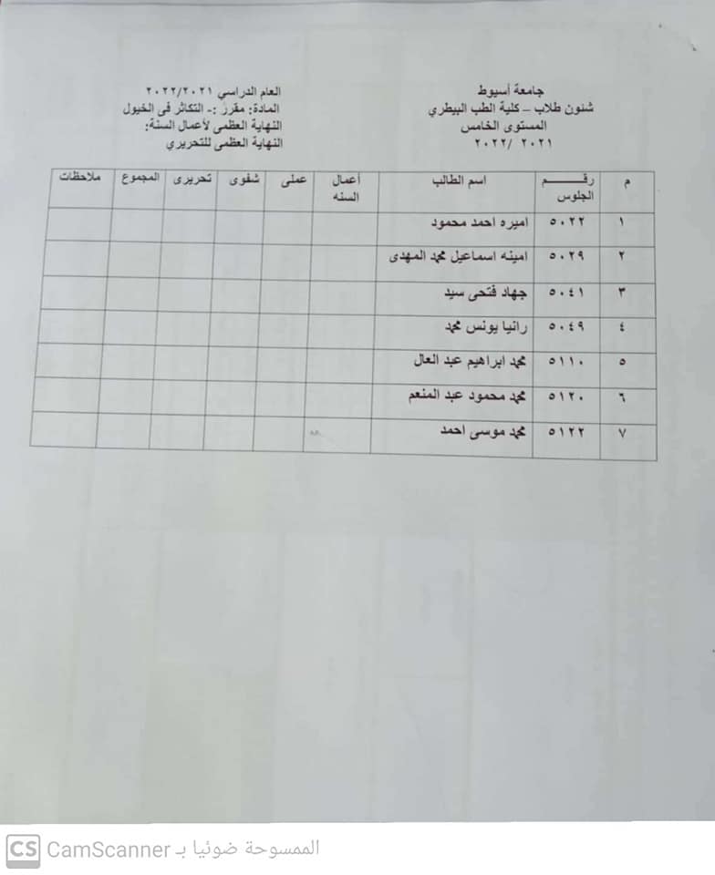
Elective courses for the fifth level 10/30/2021


Hello
veterinary students
Whoever has a problem uploading the picture of the electronic carniyah on the website link
Send me a plain background photo and the national number written in English in a private message
We are with you from 2 pm to 6 pm only
My greetings
Ms. Walaa Amer

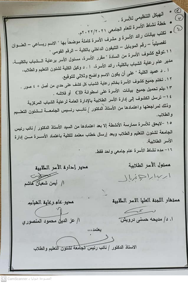
Announcement of the start of registration for student families and scientific societies for the academic year 2021-2022

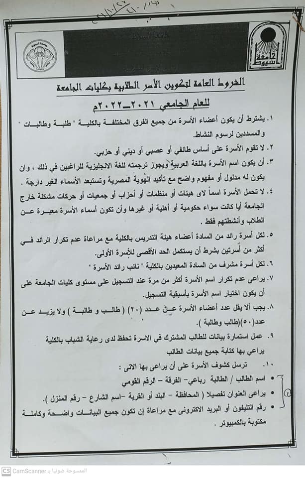
General conditions for the formation of student families in the faculties of the university for the academic year 2021-2022 AD
The adrenal gland is a vital endocrine gland that secretes many important hormones
in everyday bird life. The adrenal gland of the Japanese quail is grossly located ventromedially
the corresponding kidney and has a creamy to yellow color. The quail
gland is surrounded by a capsule and contains some ganglionic cells, and the capsule
is characterized by the presence of chromaffin cells. The adrenal gland is subdivided
into three concentric zones: subcapsular, peripheral, and central. The parenchyma
consists of interrenal tissue, chromaffin islets, and blood sinusoids. The interrenal
cells contain lipid droplets, are arranged in cords, and rest on the basement membrane.
Chromaffin cells are categorized as two types: epinephrine (E) and norepinephrine
(NE) cells. These cells contain the granules, and are characterized by the
presence of lipid droplets. In this study, the interrenal tissue was found to have a
higher proportion of chromaffin cells in quail as compared with other birds, which is
attributed to the fact that the Japanese quail is a migratory bird. Therefore, the present
investigation aims to provide a detailed study on the adrenal gland in the Japanese
quail to help physiologists understand the gland's function and the pathologist
to determine the implications for the differential diagnosis of adrenal gland tumors.
T-helper cells express CD4 as a co-receptor that binds to major histocompatibility complex class II to synchronize the immune response against upcoming threats via mediating several cytokines. We have previously reported the presence of CD4 homologues in brown trout. The study of cellular immune responses in brown trout is limited by the availability of specific antibodies. We here describe the generation of a polyclonal antibody against CD4-1 that allows for the investigation of CD4+ cells. We used this novel tool to study CD4+ cells in different tissues during viral haemorrhagic septicaemia infection (VHSV) using flow cytometric technique. Flow cytometric analyses revealed an enhanced level of surface CD4-1 expression in the infected group in major lymphoid organs and in the intestine. These results suggest an important role for the T-helper cells within the immune response against viruses, comparable to the immune response in higher vertebrates.
