settingsOrder Article Reprints Open AccessArticle Unraveling Spermatogenesis in Molly Fish (Poecilia sphenops): An Integrative Study of Testicular Ultrastructure and Immunohistochemistry

Research Abstract

Spermatogenesis in teleosts is essential for reproductive function; however, it varies considerably among species. The testis of the viviparous molly fish (Poecilia sphenops) was examined using both ultrastructural and immunohistochemical methods. The testis displays a restricted lobular type, where germ cells develop synchronously within Sertoli cell-forming cysts. Transmission electron microscopy (TEM) revealed all stages of spermatogenesis. Mature sperm are at the apex of the cysts and migrate toward the sperm ducts. Sperm duct epithelium is lined by cuboidal cells joined by tight junctions, with apical cilia and desmosomal complexes contributing to transport and structural integrity. The sperm ducts showed strong Periodic Acid–Schiff (PAS)-positive expression among negative stained spermatocysts. Centrally, a cavity serves as a storage area for spermatozoa that are organized into unencapsulated bundles known as spermatozeugmata. Sertoli cells exhibited extended cytoplasmic processes that supported developing germ cells, whereas Leydig cells occupied the interstitial tissue, contributing to hormonal regulation. Immunohistochemical labeling demonstrated strong vimentin expression in Sertoli cells and telocytes, indicating their mesenchymal origin and structural role. Calretinin expression was confined to Leydig cells and certain ductal epithelial cells, supporting its use as a marker for steroidogenic and secretory functions. These findings provide new insights into the testicular specialization of P. sphenops, highlighting key somatic–germ cell interactions, ductal adaptations, and marker expression patterns that underlie male reproductive success in viviparous fish.
Research Authors
Doaa M. Mokhtar , Giacomo Zaccone, Marialuisa Aragona , Maria Cristina Guerrera , Rasha Alonizan and Manal T. Hussein
Research Date
Research Journal
Veterinary science
Research Pages
20
Research Publisher
MDPI- Veterinary science
Research Vol
12
Research Website
https://www.mdpi.com/2306-7381/12/10/930
Research Year
2025

Evaluating Probiotic Efficacy Against Mycotoxins Threat in Semi-Dry Fermented Beef Sausage

Research Abstract

ROBIOTIC consumption is recognized as being generally safe and correlates with multiple and
valuable health benefits. Food exposure to mycotoxins is a major concern for public health
officials and regulatory authorities globally. Aflatoxins (AFLs) and ochratoxin A (OTA)
contamination of meat products can happen anywhere along the production process, from farm to
fork. The purpose of this study is to determine the concentration of AFLs and OTA residues in some
beef products and evaluate the effects of different probiotics (Lactobacillus acidophilus,
Bifidobacterium lactis and Saccharomyces cerevisiae) on AFLs and OTA in semi-dry fermented beef
sausage that has been contaminated in an experiment stored for seven days. The study found that the
AFLs were present in 86.7%, 60%, 80%, 70%, 76.7%, and 70% of the meat products under
investigation, while the OTA residues were present in 83.3%, 56.7%, 80%, 73.3%, 63.3%, and 76.7%
of the burger, minced beef, luncheon, basterma, kofta, and sausage, respectively. Burger (13.89 ±
2.62 ppb) and sausage (12.67 ± 2.37 ppb) had the greatest AFLs residues (ppb), followed by kofta
(11.38 ± 2.15 ppb) and luncheon (11.26 ± 2.72 ppb). Basterma (3.31 ± 1.85 ppb) and minced meat
(5.47 ± 1.55 ppb) had the lowest values. Luncheon had the greatest OTA residues (2.76 ± 0.43 ppb),
followed by burger (2.64 ± 0.14 ppb), sausage (2.32 ± 0.57 ppb), and kofta (1.78 ± 0.74 ppb), while
basterma (1.23 ± 0.65 ppb) and minced beef (1.56 ± 0.12 ppb) had the lowest concentrations. The
findings reveal that the levels of AFLs in some examined samples exceeded the legal limits (˂ 20
ppb), while the levels of OTA were within the acceptable range (˂ 5 ppb). The data shows a positive
association between the use of probiotics and the reduction of AFLs and OTA in all samples studied.
The results indicate that probiotics such as Lactobacillus acidophilus, Bifidobacterium lactis and
Saccharomyces cerevisiae can potentially serve as decontaminants in the food industry as well as can
replace chemical preservatives in producing organic foods and reduce the levels of mycotoxins in
beef products intended for human consumption.

Research Authors
Nady Khairy Elbarbary1*, Ashraf Abd El-malek2,3, Wageh Sobhy Darwish4, Sohaila Fathi El-Hawary5, Neveen M. Abdelmotilib6, Marwa A. Ali7 and Mohamed K. Dandrawy8
Research Date
Research Department
Research File
Research Journal
Egyptian Journal of Veterinary Sciences
Research Member
Research Pages
78-87
Research Publisher
©2025 National Information and Documentation Center (NIDOC)
Research Vol
56
Research Year
2025

Assessment of Monosodium Glutamate (MSG), sensory, physico-chemical and microbiological quality in some meat products

Research Abstract

This study aimed to assess the values of one of the harmful food additives used in food production, monosodium glutamate (MSG). A total of 60 random samples of some frozen meat products included beef burger, sausage, beef kofta and chicken nuggets (15 for each) collected from different supermarkets in Assiut City, Egypt. The samples were subjected to sensory evaluation, physico-chemical and microbiological quality. The findings revealed that the examined beef kofta samples have the lowest scores of sensory attributes, compared to beef burger and chicken nuggets samples, which recorded the highest scores. Concerning MSG, the results revealed levels were 1.415 mg/gm. in beef burger; 2.28 in sausage; 2.18 in beef kofta and 3.34 in chicken nuggets, respectively. Moreover, pH determined, and the mean values were 6.37 in beef burger; 6.22 in sausage; 6.45 in beef kofta and 6.27 in chicken nuggets, respectively. Also, the mean values for total volatile basic-nitrogen (TVB-N) (mg/100gm.) and thiobarbituric acid number (TBA) (mg MAD/kg) were 9.52 and 0.35 in beef burger; 12.13 and 0.27 in sausage; 8.87 and 0.4 in beef kofta and 11.11 and 0.23 in chicken nuggets, respectively, which within the permissible limits and all accepted according to the Egyptian standard specifications. Moreover, the bacteriological examination showed that mean values (cfu/g) of TBC and Total Y&M Count were 1.03x10⁵±5.12x10⁴ and 1.33x10⁴±7.12x10³ in beef burger; 1.43x10⁵±6.63x10⁴ and 3.73x10⁴±1.66x10⁴ in sausage; 6.75x10⁴±3.01x10⁴ and 3.2x10⁴±1.04x10⁴ in beef kofta and 5.33x10⁴ ±3.35x10⁴ and 1.98x10⁴±9.67x10³ in chicken nuggets, respectively. Furthermore, E.coli 0157:H7 identified serologically in two of both beef burger and sausage were (13.33%) and in one of chicken nuggets was (6.67%). In conclusion, application strict hygiene practices along the meat production process is important to prevent low quality products and food-borne diseases.

Research Authors
SALMA, A.A. BAKRY ; ABD-EL-MALEK, A.M. AND AMINA, M. ElRAIS
Research Date
Research Department
Research File
Research Journal
Assiut Veterinary Medical Journal
Research Member
Research Pages
447-464
Research Publisher
Faculty of Vet. Med., Assiut Uni.
Research Vol
71
Research Website
10.21608/avmj.2025.362412.1598 - Assiut University web-site: www.aun.edu.eg
Research Year
2025

The impact of dietary neem leaf on the growth and biochemical traits of rabbits

Research Abstract

Neem is a plant used both as food and in traditional medicine. Its many active components, such as Carotenoids, Saponins, Triterpenoids and Nimbidin, may render it a beneficial feed additive for rabbits. Healthy weaned rabbits from breed V-line (VL) were selected to examine the effect of neem (Azadirachta indica) on growth performance, carcass traits, morphology, and blood parameters responses. Thirty-two V-line rabbits (45 days old) were randomly assigned to four groups (n = 8 per group): a control group (G1) receiving a basal diet, and three treatment groups (G2, G3, G4) receiving the basal diet supplemented with 5%, 10%, and 15% neem leaf powder, respectively. Neem leaf supplementation had no significant effect on the rabbits' growth performance, live body weight, carcass weight, lungs and abdominal fat, dressing percentage and liver. There was a significant (P < 0.05) increase in intestine length in G4. Nevertheless, the cecum considerably shrank (P < 0.05) in G3 and G4, which might have a more negative impact on growth performance. Certain biochemical measures (albumin, globulin, triglycerides, LDL, total protein, cholesterol, glucose, AST, and ALT) did not exhibit significant variations. However, a significant (P < 0.01) drop in blood urea occurred after the higher concentration. A significant (P < 0.05) rise in HDL after neem supplementation. Histologically, the liver showed signs of hepatotoxicity in the group supplemented with neem leaves, such as abnormal hepatocytes' nuclear membranes, pyknotic nuclei, karyorrhexis and karyolysis. Additionally, the portal and central veins were congested, and a greater number of Kupffer cells were seen. In conclusion, the findings suggest that dietary neem leaf supplementation may have adverse effects on rabbit health and performance, particularly at higher concentrations.


Research Authors
Abdalla Ali , Ahmed Adawy , Zeinhom Ismaiel , Manal Hussein , Abdelraheim Attaai
Research Date
Research Journal
Scientific Reports
Research Pages
20280-20292
Research Publisher
Springer Nature
Research Rank
3.9
Research Vol
15
Research Website
https://www.nature.com/articles/s41598-025-06905-x
Research Year
2025

Sequential Development of Albino Rabbit Iris During Fetal and Postnatal Life: Histological, Ultrastructural, and Immunohistochemical Investigations

Research Abstract

The iris has several unique structures that are determined by its embryonic origin. Therefore, this research aimed to demonstrate the morphogenesis of the albino rabbit iris. The development of the rabbit iris began at mid-gestation and was completed postnatally. Consequently, we used 36 embryos and 12 neonatal rabbits. The iris originated from the tip of the optic cup. The epithelium of the iris began to differentiate on day 15 of pregnancy, forming a single layer of epithelial cells. The constrictor muscle began its differentiation on day 27 of pregnancy in the area around the pupil, while the dilator muscle differentiated on day 30 as a protrusion from the anterior epithelial cells. The stroma of the iris differentiated from the periocular mesenchyme into fibroblasts, telocytes, dendritic cells, and macrophages. The tyrosinase enzymes expressed in the albino rabbit's iris were inactive and at low levels, with melanocytes lacking melanin pigment. Vimentin immunohistochemistry expression was observed in all layers of the iris. Desmin was expressed only in the constrictor muscle and stroma. Explaining the developmental process of the albino's iris is vital for advancing our knowledge of this tissue.

Research Authors
Nagwa Ibrahim , Sara M M Eldesoky
Research Date
Research Journal
Microscopy and Microanalysis
Research Pages
ozag015
Research Publisher
Oxford University Press
Research Vol
32
Research Website
https://academic.oup.com/mam/article-abstract/32/2/ozag015/8519523?redirectedFrom=fulltext
Research Year
2026

Repeated Freeze–Thaw Cycles and Thawing Methods: Effects on Quality Attributes of Egyptian Native Chicken Breast

Research Authors
Marwa M. Abdallah & Neveen M. Abdelmotilib Nady Khairy Elbarbary, Rania S. Zaki, Ashraf Abd El-malek, Wageh S. Darwish, Marwa A. Ali
Research Date
Research Department
Research Journal
Food Biophysics
Research Member
Research Year
2026

Developmental Morphological and Histochemical Studies of the Tongue in Japanese Quails ( Coturnix japonica )

Research Abstract

The lingual apparatus of the birds was used to determine the food type of the birds. The tongue of the Japanese quail showed great plasticity in morphology, histology and function. Although little literature spoke in details about the developmental anatomy of the Japanese quail tongue and its attachment during embryogenesis and the post‐hatching period, our data were carried out on 85 healthy random specimens arranged on 45 Japanese quail embryos with ages from the 5th day of incubation to hatching day old and 40 quail chicks with the age of 7, 14, 30 and 60 days old. Those investigations found that the primordium of the tongue arose from the oropharyngeal floor primordium as a clear elongated projection supported by basihyale at the 5th and 6th days old of the embryonated egg. The tongue primordium was attached by a thin layer of epithelium (frenulum linguae primordium). With age advancing, the tongue took different shapes, starting from a ‘U’ shape to a triangular elongated shape. Consequently, its parts were fully developed into a lingual apex that was supported by the paraglossale apical process and paraglossale corpus and the lingual body supported by both the posterior part of paraglossale and basihyale. Besides that, (ala linguae) primordia became more prominent as two oval projections with caudal serrated borders at the 10‐day‐old embryo which were supported by posterior processes of os paraglossale. But at 13 days old, the transverse papillary crest primordium began to appear, which consisted of caudally directed finger‐like papillae. Those papillae were developmentally variable in number, shape and length. The lingual muscle primordia could be noticed: ceratoglossus, hypoglossus anterior and genioglossus muscles at 5, 6 and 7 days old of incubation, respectively. However, the genioglossus muscle showed a degenerative process at the 11‐day‐old embryo. Lingual glands were compound tubular and tubulo‐alveolar end pieces, which its primordia appeared at the 7 and 8‐day‐old embryo. Histochemically, the glands showed different reactions to AB and PAS, and combined stains, which the developmental stains reaction ended with a slightly alcinophilic reaction.

Research Authors
Mahmoud Osman Khalifa, Wafaa Gaber, Abdelmohaimen Mostafa Saleh
Research Date
Research Journal
Anatomia Histologia Embryologia
Research Pages
1-26
Research Publisher
WILEY
Research Vol
55(2)
Research Website
DOI:10.1111/ahe.70083
Research Year
2026

Most Beautiful Ramadan Square Competition 2

Under the patronage of
Professor Dr. Ahmed El-Menshawy, President of the University
Professor Dr. Ahmed Abdel-Mawla, Vice President for Education and Student Affairs
Professor Dr. Enas Ahmed Abdel-Hafez, Dean of the Faculty
Professor Dr. Khaled Mohamed Ahmed Hassanin, Vice Dean for Education and Student Affairs
Professor Dr. Madiha Darwish, General Coordinator of Student Activities at the University
And under the supervision of Dr. Ahmed Thabet Abdel-Awad, Director of the Student Welfare Department
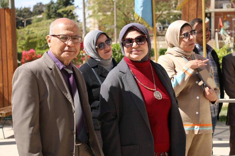
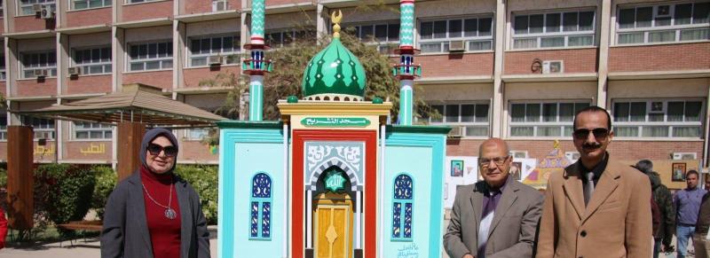
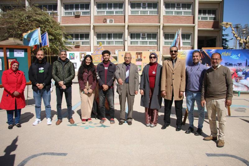
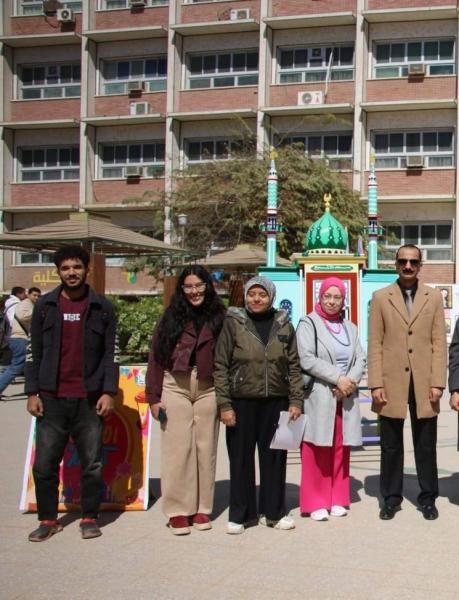
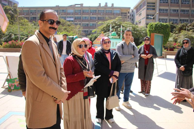
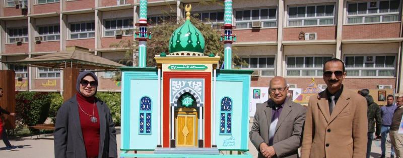
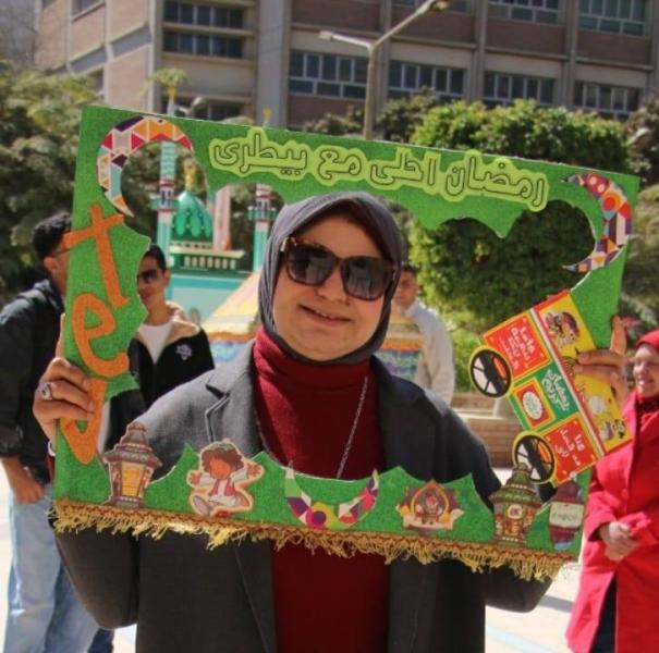
The Technical Committee of the Student Welfare Department at the Faculty of Veterinary Medicine participated in the "Most Beautiful Ramadan Courtyard 2" competition. The evaluation was attended by Professor Dr. [Name of Dean], Professor Dr. [Name of Vice Dean], Professor Dr. [Name of Vice Dean], Professor Dr. Mohamed Ahmed Alam El-Din, Supervisor of the Anatomy Department Museums, and an evaluation committee from the Arts Department of the General Administration for Student Welfare at Assiut University, on Sunday, March 1, 2026.

All praised the high standard of the Ramadan displays in the courtyard of the Faculty of Veterinary Medicine.

1

Art exhibition by student Mohamed Refaat (fourth year) and student Jessica Raafat Fayek (first year)

0

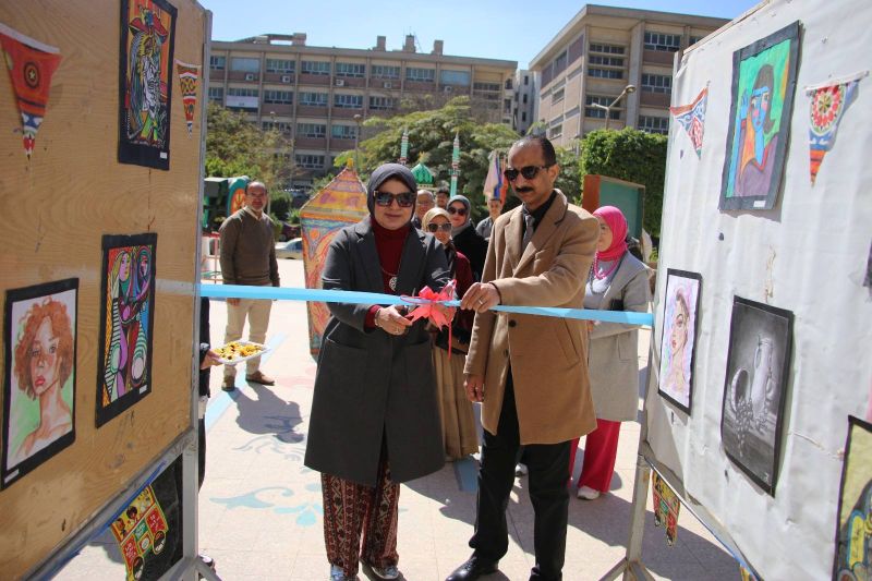
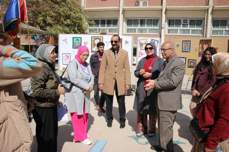
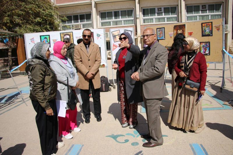
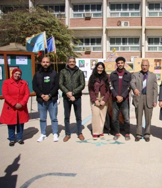
Under the patronage of Professor Dr. Ahmed El-Menshawy, President of the University; Professor Dr. Ahmed Abdel-Mawla, Vice President for Education and Student Affairs; Professor Dr. Enas Ahmed Abdel-Hafez, Dean of the Faculty; Professor Dr. Khaled Mohamed Ahmed Hassanin, Vice Dean for Education and Student Affairs; and Professor Dr. Madiha Darwish, General Coordinator of Student Activities at the University; and under the supervision of Dr. Ahmed Thabet Abdel-Awad, Director of the Student Welfare Department, the Technical Committee of the Student Welfare Department at the Faculty of Veterinary Medicine organized an art exhibition for student Mohamed Refaat (fourth year) and student Jessica Raafat Fayek (first year). The exhibition was inaugurated by Professor Dr. the Dean of the Faculty and Professor Dr. the Vice Dean for Education and Student Affairs, in the presence of a select group of supervisors of fine arts from the Arts Department of the General Administration for Student Welfare at Assiut University. The exhibition opened on Sunday, March 1, 2026, and continued until Tuesday, March 3, 2026, in the Faculty grounds.

All attendees praised the high quality of the artwork. The art in the exhibition

215
Subscribe to